Anatomy of the Knee Joint
The human knee joint is a complex hinge joint responsible for a wide range of movements. Understanding its intricate structure is crucial for comprehending how it functions and the potential for injuries. A key element involves the femur, tibia, and patella—the thigh bone, shinbone, and kneecap, respectively. These bones articulate to form the knee joint’s primary structure. The knee joint’s intricate design includes the menisci, crucial cartilage structures that act as shock absorbers. A clear diagram of a knee can illustrate the arrangement of these bones. Moreover, ligaments and tendons play essential roles in providing stability and support to the knee joint. Crucial ligaments, such as the anterior cruciate ligament (ACL) and posterior cruciate ligament (PCL), help maintain the knee’s alignment. The menisci further enhance stability by cushioning and distributing forces during movement. Proper functioning of the knee hinges on the precise alignment and proper interplay of these structural elements. A detailed diagram of a knee joint can clarify the arrangement of these components. The patella, or kneecap, slides within a groove of the femur and facilitates knee extension.
The knee joint’s stability also depends on tendons, such as the patellar tendon and quadriceps tendon. These tendons connect muscles to the bones and contribute to the knee’s overall function. A diagram of a knee’s tendons can provide a clear understanding. Ligaments provide crucial stability by connecting bones, whereas tendons connect muscles to bones, enabling movement. A clear diagram of a knee joint can aid in comprehension by visualizing the interplay of these structural components. The intricate interplay of these structures ensures the knee’s functionality and the maintenance of its structural integrity.
The menisci, specialized cartilage pieces, cushion and distribute force across the knee joint during movement. A diagram of the knee joint, emphasizing the menisci’s placement, can greatly aid in understanding their crucial role. The knee’s unique structure allows a broad range of movements, from flexion and extension to rotation. The interplay between bones, ligaments, and tendons, along with supportive structures like the menisci, is pivotal in ensuring the knee’s stability and mobility. A comprehensive diagram of a knee’s structure would showcase the detailed arrangement of all these components.
Types of Knee Movements and Their Mechanics
The knee joint, a complex structure, permits several types of movement crucial for daily activities and physical performance. These movements, facilitated by the interplay of bones, ligaments, muscles, and tendons, include flexion, extension, and, to a lesser extent, rotation and adduction/abduction. Flexion refers to bending the knee, decreasing the angle between the thigh and lower leg. Extension is the straightening of the knee, increasing the angle. A diagram of a knee illustrating these movements would clearly show the changing angles between the femur and tibia.
Rotation, while limited in a fully extended knee, becomes more pronounced when the knee is flexed. This allows for subtle twisting movements, essential for activities like turning and pivoting. Adduction involves moving the lower leg towards the midline of the body, while abduction moves it away. These lateral movements are less significant than flexion and extension but contribute to overall knee functionality. Examining a diagram of a knee during these movements reveals the complex interplay of the various structures. Understanding the mechanics of these movements is vital, especially for athletes and individuals engaging in strenuous physical activity. Proper technique minimizes stress on the joint. A detailed diagram of a knee will show the bones moving in relation to each other and the ligaments’ role in limiting excessive range of motion.
The patella, or kneecap, plays a key role in knee mechanics. It acts as a pulley, improving the efficiency of the quadriceps muscle group during knee extension. The menisci, crescent-shaped cartilages, act as shock absorbers, distributing forces evenly across the joint surfaces. Ligaments and tendons provide crucial stability, preventing excessive or unwanted movements. A diagram of a knee highlighting these elements is essential to fully grasp the mechanics of knee movement. By examining such a diagram, one can appreciate the coordinated action of these structures. This understanding helps in preventing injuries and promoting optimal knee health.
Ligaments and Tendons: The Knee’s Supporting Structures
The knee joint relies on a complex network of ligaments and tendons for stability and support. These structures work together to prevent excessive movement and protect the joint from injury. A diagram of a knee clearly shows the crucial roles these components play. The four major ligaments are the anterior cruciate ligament (ACL), posterior cruciate ligament (PCL), medial collateral ligament (MCL), and lateral collateral ligament (LCL). The ACL and PCL are intracapsular ligaments, meaning they are located within the joint capsule. The ACL prevents the tibia from sliding forward under the femur, while the PCL prevents the tibia from sliding backward. The MCL and LCL are extracapsular ligaments, providing medial and lateral stability respectively. Damage to any of these ligaments can result in instability and pain. A diagram of a knee illustrating these ligaments is essential for understanding their precise locations and functions.
Tendons connect muscles to bones, and several crucial tendons contribute to knee function. The patellar tendon connects the patella (kneecap) to the tibia, transmitting forces generated by the quadriceps muscles during knee extension. The quadriceps tendon connects the quadriceps muscles to the patella. These tendons play a vital role in the powerful extension of the knee. Proper functioning of both the patellar and quadriceps tendons is crucial for activities involving jumping, running, and even simple walking. A detailed diagram of a knee highlighting these tendons and their connections to muscles helps visualize their significant impact on knee movement. Understanding the interaction between ligaments and tendons, shown clearly in a diagram of a knee, provides a comprehensive understanding of knee joint mechanics. The stability and functionality of the knee greatly depend on these structures working in harmony.
A clear diagram of a knee is invaluable for understanding the intricate interplay of ligaments and tendons. This diagram should illustrate the relative positions of the ACL, PCL, MCL, LCL, patellar tendon, and quadriceps tendon. It should also show how these structures are interwoven to provide comprehensive stability and enable smooth knee movement. Consider the forces acting on the knee during various activities, a diagram of a knee helps visualize how these structures absorb and distribute these forces, preventing injury. Detailed diagrams available online provide different perspectives, offering a complete picture of these essential knee components. Studying these visuals clarifies how specific injuries might affect movement and stability. Furthermore, understanding the location and function of each ligament and tendon facilitates better injury prevention and rehabilitation strategies. By providing a visual understanding of the knee’s support structures, a diagram of a knee serves as a powerful educational tool.
Muscles and their Role in Knee Stability
The knee joint relies on a complex interplay of muscles for stability and movement. Understanding these muscles is crucial for comprehending both normal knee function and the potential causes of injury. A diagram of a knee clearly illustrates the key muscle groups involved. The quadriceps group, located on the front of the thigh, is responsible for extending the knee. This group comprises four muscles: rectus femoris, vastus lateralis, vastus medialis, and vastus intermedius. Their coordinated action straightens the leg. The rectus femoris also flexes the hip. Examining a diagram of a knee helps visualize these muscle attachments and their actions.
On the back of the thigh, the hamstring group—biceps femoris, semitendinosus, and semimembranosus—flexes the knee and extends the hip. These muscles are vital for activities like walking, running, and jumping. They also play a crucial role in knee stability, especially during movements involving bending and straightening. A clear diagram of a knee helps visualize the location and actions of these opposing muscle groups. Weakness or imbalance in either the quadriceps or hamstrings can increase the risk of knee injury. The gastrocnemius and soleus muscles, located in the calf, also contribute to knee flexion, although their primary function is ankle plantarflexion. Understanding their role provides a complete picture of knee biomechanics. A detailed diagram of a knee will help understand the integrated workings of all these muscles.
Proper muscle balance and strength are essential for knee health. Exercises that target both the quadriceps and hamstrings are crucial for maintaining stability and preventing injuries. Understanding muscle function, as revealed by a diagram of a knee, allows for targeted strengthening and injury prevention strategies. Regular exercise and balanced muscle development minimize strain and risk of knee problems. A thorough understanding of the anatomy, as shown in a diagram of a knee, contributes to better care and injury prevention.
How to Avoid Knee Pain: Prevention Strategies
Maintaining knee health involves a multifaceted approach. Regular exercise plays a crucial role. However, proper techniques are essential. Incorrect form can strain the knee joint. A good warm-up routine prepares the muscles and ligaments. This reduces the risk of injury. A diagram of a knee clearly shows the interconnectedness of these structures. Understanding this relationship is vital for effective prevention. Remember to gradually increase intensity and duration to avoid overexertion. Incorporate a variety of exercises targeting different muscle groups. This ensures balanced strength and flexibility. For instance, strengthening the quadriceps and hamstrings stabilizes the knee. These muscles are essential for supporting the knee joint, as shown in any diagram of a knee. This balanced approach significantly reduces the risk of injury and subsequent pain.
Body weight significantly impacts knee health. Excess weight puts added stress on the knee joint. This increases the risk of osteoarthritis and other knee problems. Maintaining a healthy weight through balanced diet and regular physical activity is crucial. A proper diet supports overall health, reducing the strain on the knee joint. Remember to choose footwear suitable for your activities. Proper shoes provide cushioning and support, reducing the impact on the knees. Selecting shoes that fit correctly and offer adequate support prevents further issues. A diagram of a knee shows the importance of proper alignment. Shoes influence this alignment. This is crucial for injury prevention. This includes selecting shoes with good arch support and adequate cushioning. These features minimize stress on the knee joint during movement and impact.
Specific exercises can greatly strengthen the muscles surrounding the knee. These exercises improve stability and reduce the risk of injury. Simple exercises like squats, lunges, and hamstring curls strengthen key muscles. These muscles provide crucial support. They are critical for maintaining knee stability, as illustrated in a diagram of a knee. Always consult a healthcare professional before starting a new exercise program. They can create a plan tailored to your specific needs and fitness level. Proper form and gradual progression are key. This prevents further injury. Remember, a proactive approach to knee health ensures long-term well-being. Understanding the anatomy, as depicted in a diagram of a knee, empowers individuals to make informed choices. This leads to improved knee health and reduces the likelihood of experiencing pain.
Common Knee Injuries: Identifying the Symptoms
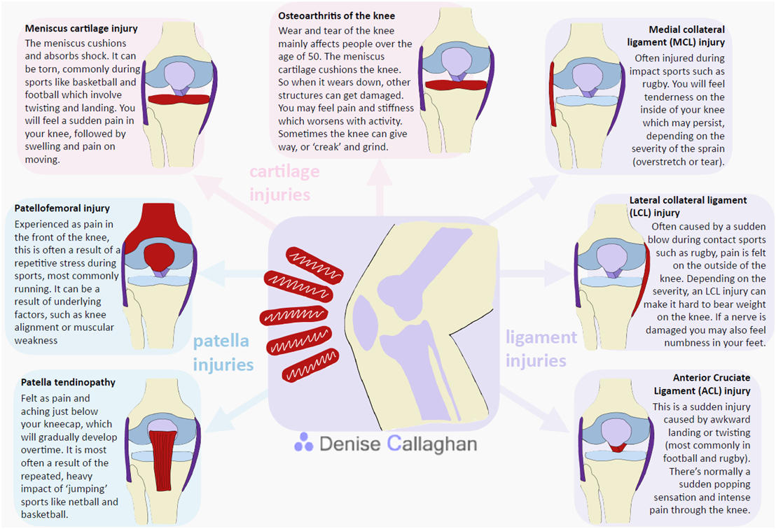
The knee, a complex joint, is susceptible to various injuries. Understanding these injuries and their symptoms is crucial for timely intervention. A diagram of a knee clearly shows the structures involved, making it easier to comprehend the impact of different injuries. Among the most prevalent are ACL tears, meniscus tears, patellofemoral pain syndrome, and bursitis. ACL tears, often occurring during sports, involve a rupture of the anterior cruciate ligament. Symptoms include immediate pain, swelling, instability, and a popping sensation. A diagram of a knee illustrating the ACL’s location helps visualize the injury. Meniscus tears, usually caused by twisting or forceful impacts, affect the cartilage pads within the knee. Symptoms include pain, swelling, stiffness, locking, and difficulty bearing weight. A comprehensive diagram of a knee will show the location of the menisci and their function.
Patellofemoral pain syndrome, commonly known as runner’s knee, involves pain around the kneecap. This condition often arises from overuse, muscle imbalances, or structural abnormalities. Symptoms include pain behind or around the kneecap, especially during activities involving bending the knee. Pain may increase after prolonged sitting or climbing stairs. A diagram of a knee showing the patella and surrounding structures illustrates the source of the pain. Bursitis, an inflammation of the bursae (fluid-filled sacs cushioning the knee joint), causes pain and swelling. Repetitive movements or direct trauma may trigger this condition. Symptoms include localized pain, tenderness, and swelling. A clear diagram of a knee can pinpoint the location of the bursae and their role in reducing friction.
Accurate diagnosis is paramount for effective treatment. A thorough medical evaluation, often including physical examination and imaging studies, will confirm the specific injury. Understanding the underlying cause and the precise location of the damage—as visualized in a diagram of a knee—is critical for developing an appropriate treatment plan. Early intervention usually leads to better outcomes. Early diagnosis and treatment minimize complications and aid in the restoration of full function. Remember, a diagram of a knee helps illustrate these injuries, making the process of understanding and treating them much clearer.
What to Do If You Suspect a Knee Injury
Suspecting a knee injury requires immediate attention. Ignoring symptoms can lead to worsening conditions. A proper diagnosis is crucial for effective treatment. Seek medical attention promptly. A doctor can accurately assess the extent of the damage. They will perform a physical examination. They may also order imaging tests, such as X-rays or MRIs. These tests provide a detailed view of the knee joint. A clear diagram of a knee, from these tests, helps in identifying the specific injury. Early intervention helps to prevent long-term problems. This is especially true for ligament or meniscus tears.
During your consultation, describe your symptoms clearly. Note when the injury occurred. Explain the mechanism of the injury. Details like the direction of force or a twisting motion are helpful. Be prepared to answer questions about your medical history. Your doctor may ask about any previous knee problems. The severity of your pain and any limitations in movement should also be discussed. Based on the assessment, your doctor will recommend a course of action. This could range from rest and physical therapy to surgery. Remember that proper diagnosis is key. Self-treatment can be dangerous. It can delay proper healing and increase the risk of complications. Trust the expertise of a medical professional.
Following your doctor’s instructions is vital. Physical therapy plays a significant role in recovery. It helps restore strength and range of motion. A well-structured rehabilitation program will guide you back to your normal activities. A thorough understanding of knee anatomy, including a clear diagram of a knee joint, aids in recovery. Understanding the underlying structures enables better compliance with the treatment plan. This leads to faster healing and a reduced risk of re-injury. Remember, your doctor is the best resource for guidance. They will help you navigate your recovery journey. Do not hesitate to contact your physician if any new or concerning symptoms arise. A clear diagram of a knee can be instrumental in understanding the diagnosis and rehabilitation process.
Additional Resources for Knee Pain Relief
Understanding the complexities of the knee joint often requires more than one resource. To further enhance your knowledge, consider exploring reputable online platforms dedicated to musculoskeletal health. Websites from organizations like the American Academy of Orthopaedic Surgeons (AAOS) and the Arthritis Foundation provide evidence-based information on knee anatomy, common injuries, and treatment options. These sites frequently feature detailed diagrams of a knee, illustrating the intricate network of bones, ligaments, and tendons. They often offer interactive tools and visual aids, making complex anatomical concepts easier to grasp. A thorough understanding, aided by a diagram of a knee, empowers individuals to make informed decisions about their knee health.
Beyond online resources, physical therapy plays a crucial role in managing knee pain and improving function. Physical therapists assess individual needs and design tailored exercise programs. These programs strengthen supporting muscles, improve flexibility, and enhance overall knee stability. A diagram of a knee can be used as a guide during therapy sessions, helping to visualize the targeted muscles and movements. The therapists can adapt exercises according to your progress, providing ongoing support and guidance for long-term knee health. Remember to always consult with your doctor or physical therapist before starting any new exercise regimen.
Books on anatomy and kinesiology can provide a deeper dive into the mechanics of the knee joint. These often include detailed illustrations and diagrams of a knee, clarifying the relationships between different structures. Furthermore, seeking a second opinion from a specialist, such as an orthopedic surgeon, can be beneficial for complex cases or persistent symptoms. These experts can offer specialized insights and advanced diagnostic tools, ensuring you receive the most appropriate and effective care. Remember, proactive management and a comprehensive understanding, aided by studying a diagram of a knee and other resources, are key to maintaining healthy knees throughout life.


